Multiscale Assessments Research (MAR) Core
Multimodal Animal Imaging Lab
Mary Boggs, PhD
Dr. Mary Boggs completed her PhD at the University of Delaware in the Department of Biological Sciences under the direction of Dr. Randall Duncan from Biological Sciences and Dr. Tom Beebe, Jr. from the Department of Chemistry and Biochemistry in 2011. Her graduate work focused on developing bone/nerve cell co-culturing methods and identifying mechanisms of cell-cell communication between these cells. Post graduation, she stayed on as a Research Associate in the Duncan Lab and was involved in a variety of projects aimed to understand biological molecules involved in bone and cartilage responses to mechanical forces at both the cell and organismal level. Before joining the DCMR, Dr. Boggs served as the Associate Director of DBI Research at the Delaware Biotechnology Institute. In this role she provided K-12 STEM education opportunities and training and provided oversight to the shared instrument facilities at the DBI. Additionally, she served as an Associate Director for the Chemistry/Biology Interface Program where she provided insight and advice to student progress and engaged students in outreach activities and resilience training. As the Lead Scientist for the DCMR, Dr. Boggs is excited to provide her expertise and technical support for those using the DCMR Core Instrumentation.

Services offered:
- Analysis Assistance
- Assay/Protocol Development
- Consultation/Training
- Project Development
Multimodal Animal Imaging Center Equipment:
Agilent BioTek Cytation C10 Confocal Imaging Reader
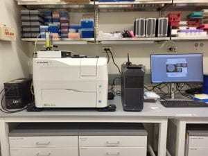 The Cytation C10 is a confocal imaging reader that combines automated confocal and widefield microscopy with multimode plate reading. The system is outfitted with a Hamamatsu Orca sCMOS, 16-bit grayscale camera for use in confocal and widefield imaging. For confocal imaging, users can select between 60μm and 40μm Nipkow spinning discs as well as 6 different laser lines. Available features for confocal and widefield imaging include CY5, DAPI, GFP, RFP, YFP (widefield only) and TRITC filters as well as 1.25, 4, 10, 20, 40 and 60x objectives (confocal is only available for 20, 40 and 60x). The system provides imaging modes including single and multi-color, time lapse, montage, z-stacking and z-stack montage. Detection modes include UV-Vis absorbance, fluorescence intensity and luminescence. The Cytation C10 accepts multi-well microplates ranging from 6 to 1536 wells for imaging and 6 to 384 wells for detection. Other supported labware includes microscope slides, petri and cell culture dishes, cell culture T25 flasks and hemocytometer. To maintain proper environment during time lapse or kinetic studies, temperature can be controlled from room temperature up to 45ºC and CO2 can be controlled from 0-20%.
Brüker SkyScan 1276 Micro-CT
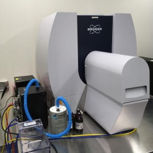 The SkyScan 1276 is a high performance, stand-alone, fast, desktop in vivo micro-CT with continuously variable magnification for scanning small laboratory animals (mice, rats, hamster, etc.) and biological samples.
Description: The Brüker SkyScan 1276 Micro-CT features a combination of high resolution, big image size, round/spiral scanning, reconstruction, and low dose imaging. The image field of view (up to 80 mm wide and more than 300 mm long) allows full body mouse and rat scanning. The variable magnification allows scanning bone and tissue samples with high spatial resolution down to a 2.8 µm pixel size. Variable X-ray energy combined with a range of filters ensures optimal image quality for diverse research applications from lung tissue to bone with metal implants. Further, the SkyScan 1276 in vivo micro-CT administers low radiation dose to the animals allowing multiple scans in longitudinal pre-clinical studies without the risk of unwanted radiation-induced side effects. The system can perform scanning with continuous gantry rotation and in step- and-shoot mode with the fastest scanning cycle of 3.9 sec.
The SkyScan 1276 allows reconstructing non-invasively any cross section(s) through the animal body with possibilities to convert reconstructed datasets into realistic 3D-images and to calculate internal morphological parameters including specific bone structural parameters.
Features:
- 3.9 second shortest scanning cycle
- 2.8 micron highest nominal resolution
- Maintenance-free 20-100 kV micro-focus X-ray source, automatic 6-position filter changer
- 11 Mp cooled X-ray camera
- Continuously variable magnification with smallest pixel size of 2.8 micron
- Possibility to resolve object details of 5-6µm with more than 10% contrast
- Scanning in circular or spiral (helical) trajectory
- GPU-accelerated and world’s fastest InstaRecon® 3D reconstruction
- Full body mouse and rat scanning, 80mm scanning diameter, >300mm scanning length,
- Step-and-shoot and continuous gantry rotation with shortest scanning cycle of 3.9 sec.
- Integrated physiological monitoring (breathing, movement detection, ECG) for time-resolved 4D microtomography
- Software for 2D/3D image analysis, bone morphology and realistic visualization
- On-screen dose meter to estimate accumulated dose and dose rate
- Export results to 3D printers to create magnified physical copy of scanned objects
Cellink BioX6 Bioprinter
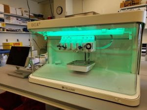 The BioX6 Bioprinter is a high-throughput bioprinter for cell culturing and biofabrication. The BioX6 provides 6 printheads to print more complex structure and physiologically relevant models. The printhead temperatures can range from 4-250ºC with a printbed temperature of 4 to 65ºC allowing for precise control when working with temperature-sensitive materials. The bioprinter is enclosed in a benchtop biosafety cabinet with a HEPA filter and 4 build-in UV modules to maintain sterility around the print area.
FujiFilm VisualSonics Vevo F2 LAZR-X Photoacoustic Ultrasound
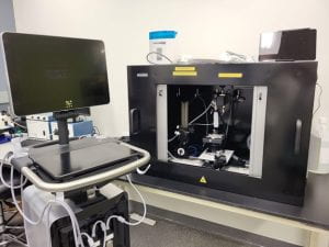 The Vevo F2 LAZR-X Photoacoustic Ultrasound is a multimodal imaging system that provides high resolution, high frequency ultrasound imaging of rodent models including mouse and rat. Ultrasound imaging depths can be up to 9cm with lower frequency transducers, while resolution can be increased (down to 30μm) using higher-frequency transducers. The added LAZR-X allows users to visualize real-time oxygen saturation and molecular imaging, co-registered with high resolution anatomy. Available ultrasound transducers include UHF29x, UHF46x and UHF71x. Photoacoustic imaging is available using the UHF29x and the UHF46x transducers.
IVIS Lumina III In Vivo Imaging System
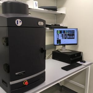 The IVIS ® Lumina Series III brings together years of leading optical imaging technologies into one easy to use and exquisitely sensitive bench-top system. The IVIS Lumina III is capable of imaging both fluorescent and bioluminescent reporters. The system is equipped with up to 26 filter sets that can be used to image reporters that emit from green to near-infrared. Superior spectral unmixing can be achieved by Lumina III’s optional high resolution short cut off filters.
Features:
- Exquisite sensitivity in bioluminescence
- Full fluorescence tunability through the NIR spectrum
- Compute Pure Spectrum spectral unmixing for ultimate fluorescence sensitivity
- Dedicated XGI-8 Gas Anesthesia System
Location: CBBI (Center for Biomedical and Brain Imaging) 244A
Contact person: Dr. Mary Boggs
Scanco Medicine 35 micro CT
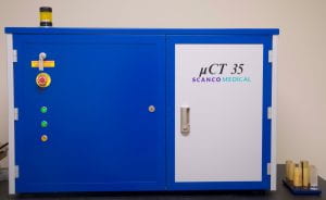
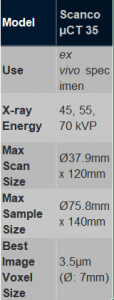 The Scanco 35 microCT system is used to exam diverse materials including bone and other hard tissues as well as medical devices. This system can be used to assess trabecular bone structure from tibia, femor and vertebrae harvested from rats and mice.
Wacom Cintiq Pro 24 touch
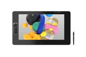
A touch tablet with wireless pen for precision data segmentation.
Workstations
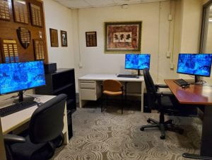 The DCMR supports a set of computational workstations in 203 Spencer Lab (2 units), 244 CBBI (1 unit) and 115 Wolf hall (3 units). Each set of workstations provide software for offline data analysis for equipment related to multimodal imaging including the Bruker Skyscan softwares and Vevo Lab, and other softwares such as ImageJ, MATLAB, Python and the Materialise Mimics software suite for segmentation and FEA.
Histology Lab
Charles Riley, AS, BS, HTL (ASCP)
Charles graduated from Delaware Technical Community College in May 2014 with his associates degree in Histotechnology. Obtained ASCP HT board certification two weeks after graduating. Charles worked at Doctors Pathology services from July 2014 until April of 2021. During his tenure there, Charles became a certified trained MOHS technician, managed the Anatomic pathology division (histology and grossing departments), and obtained his Bachelor’s degree from Wilmington University in Allied Health Management. Charles became ASCP HTL certified in 2017. Charles has advanced experience with training new users, tissue processing techniques, microtomy, cryotomy, and H&E and immunohistochemistry (IHC) staining and troubleshooting.
Services offered:
- Analysis assistance
- Assay/Protocol Development
- Consultation/Training
- Equipment education
- Histology techniques education
- Project development
- Frozen section microtomy
- H&E staining
- IHC testing (manual)
- IHC testing (automatic)
- Special stain studies (automatic)
- Sample processing
- Slide coverslipping
- Special stain studies (manual)
- Tissue embedding (paraffin)
- Tissue Microtomy (paraffin)
- Tissue processing
Coming soon:
- Tissue embedding (resin)
- Tissue Microtomy (resin)
Histology Lab Equipment:
Avantik QS12 Cryostat
The Avantik QS12 Cryostat provides the following features: 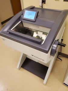
- Touch interface for control of all cryostat functions
- Unique “dovetail” bladeholder base design to provide enhanced stability and prevent lateral movement or vibration
- Fully adjustable, extra-wide anti-roll glass
- Automatic specimen retraction
- Precise trim thickness, section thickness and sectioning depth measurements
- Memory function allowing the operator to record the last position of the specimen relative to the blade before removing it. The specimen can be automatically returned to the same position if additional sectioning is required.
ESPO Thermal Transfer Slide Printer
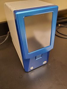 The ESPO Thermal Transfer Slide Printer provides direct labeling to slide, creating a more efficient work flow and minimizing errors.
The system features include:
- Fully integrated color touch-screen display
- Two 100 slide cartridges
- Single-slide front loading
- Barcode capabilities
- Solvent resistant: water, alcohol, xylene
Slide Compatibility
- Print speed: 3 seconds per slide, 18 slides per minute
- Print direction (in degrees): 0, 90, 180, 270
- Slide size: 25mmx75mmx1mm; 26mmx76mmx1mm
- Frost size: 15mm, 20mm, 25mm
HistoDream Embedding Workstation
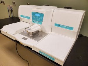 The Milestone HistoDream Embedding Workstation is composed of 3 separate units – the dispensing module, thermal module and cold embedding module. The Dispensing module is compatible with all cassette types and has 2 built-in paraffin trimmers, heated forcep wells and programmable heating. The Thermal module can store up to 400 molds and provides programmable heating. The Cold Embedding module provides even temperature distribution over the entire cooling surface, an can operate in the temperature range of 0 to -12°C.
The HistoDream Embedding Workstation is used for the following operations:
- Dissolve solid wax for specimen embedding and to keep it melted at the required temperature
- Fill the molds where the tissue specimens have been placed with wax
- Heat and keep the temperatures of the moldes and tweezers
IsoMet Low Speed Saw
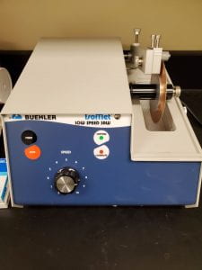 The IsoMet Low Speed Saw is designed for cutting of various types of materials with minimal deformation. The low speed cutter is targeted for delicates parts by only using gravity fed force.
Features:
- Gravity fed cutting force to reduce deformation of delicate samples
- Cut speeds of 0-300 RPM
- Capable of cutting a variety of materials including biomaterials.
- Capable of cutting ±5μm precision via a manual micrometer
Leica CM 1100 Tabletop Cryostat
 The Leica CM1100 is a portable compact cryostat for rapid freezing and manual sectioning of tissue specimens.
Features
- Portable bench-top cryostat
- Compact unit, providing maximum ease of use at a total weight of 50 kg
- Spacious, easy-to-clean cryochamber made out of stainless steel
- CE disposable blade holder with lateral adjustment and glass anti-roll plate
- Quick-freeze shelf
- Two section waste trays
- Microtome with specimen cylinder slot cover – ideal for spray disinfection
- Automatic and manual hot gas defrosting
- CFC-free refrigerants and insulating foams.
- Connection for 12-volt car battery
Technical Specifications:
- Section thickness adjustment: 0 to 20 μm
- Maximum specimen size: 25 mm ø
- Total horizontal specimen advance: 10 mm
- Vertical specimen stroke: 46 mm
- Dimensions (W x H x D): 570 x 380 x 777 mm
- Dimensions cryocabinet (W x H x D): 265 x 220 x 400 mm
- Weight (including microtome): 110 lbs.
Leica RM2125 Microtome
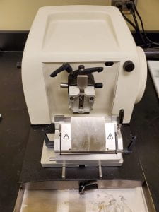
The Leica RM2125 Microtome is a manual microtome that can provide sectioning thickness of 0.5-60μm and trimming thickness of 10 or 50μm
Leica RM2155 Microtome
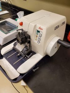 The Leica RM2155 is a reliable rotary microtome designed for fully motorized sectioning of both paraffin and resin-embedded specimens. Offering a broad application spectrum in histology laboratories, the RM2155 also has applications in industrial materials and quality control. The Leica microtome’s two-in-one design concept allows motorized as well as manual sectioning, and provides reproducible quality sections.
The RM2155 sectioning speed can be adjusted to cut hard materials as well as paraffin-embedded specimens at high throughput without affecting sectioning quality. This microtome is designed for users who require a fully motorized/electronic microtome for their laboratory needs, making it an ideal instrument for fundamental sectioning of standard and non-standard materials.
The fully motorized Leica RM2155 microtome can cut sections from 0.5 to 60 microns with an automatic motorized coarse feed as well as an adjustable hand wheel for speed control and selectable cutting modes. The knife holder comes with new clamping plates, ensuring the user accurate, non-erratic sectioning.
PiSmart Single Hopper Plus Cassette Printer
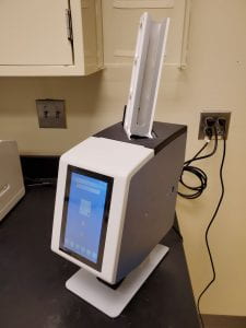 Designed for on-demand printing, the PiSmart Single Plus can deliver the first printed cassette in as little as 3 seconds. Users have the ability to bypass the hopper and manually drop in a different type of cassette.
Key Features:
- Delivers printed cassettes in 3-5 seconds
- High-quality print finish
- Full-color touch screen
- Low printing error rate
Tissue-Tek Film 4740 Coverslipper
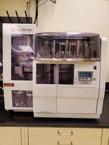 The Tissue-tek Film Automated Coverslipper utilizes a xylene-activated film to apply proper coverage to samples. Film coverslipped slides have fewer artifacts compared to glass coverslipped slides. The system features include:
- Quick drying
- Barcode scanning
- Holding capacity of 240 slides
- Can accommodate coverslip lengths of 45, 50, 55 or 60mm
Tissue-Tek Genie Advanced Staining System
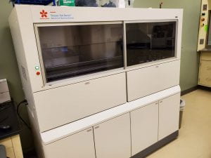 The Tissue-Tek Genie Advanced Staining System is a fully automated stainer for application in immunohistochemistry and in situ hybridization.
Features:
- Turnaround time of 2-3hr for IHC including dewaxing, antigen retrieval and counterstaining
- Throughput of up to 90 IHC slides in 8 hours
- Temperature range of 10-98°C
- Slides requirements: 25x75mm or 26x76mm positively charged slides
Invitrogen EVOS M7000
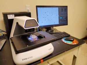
The Invitrogen EVOS M7000 is a high-performance, fully automated, inverted, multi-channel fluorescence and transmitted light imaging system. The system is designed for basic image acquisition or automated routines for time-lapse live cell imaging, area scanning in time lapse, Z-stack, tiling and stitching.
Features:
-
4, 10 and 20X objectives
-
LED cubes for DAPI, GFP, RFP and Cy5
-
Image analysis capabilities for cell segmentation and quantification
Leica ST5010 Autostainer XL
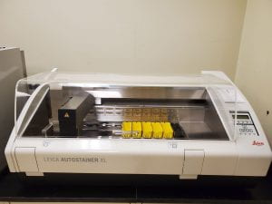
The Leica ST5010 Autostainer XL provides reproducible, consistent high-quality staining and increased workload throughput in comparison to manual staining.
Features:
-
Ability to store and run up to 15 user-defined staining protocols
-
High specimen throughput with up to 11 racks and 30 slides at a time
-
Easy-to-use software
Leica ASP6025 S Tissue Processor
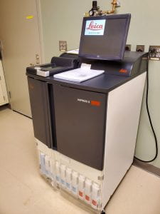 The Leica ASP6025 S Tissue Processor provides end-users with optimal tissue processing speed and quality.
Features:
-
Advanced magnetic stirrer technology to optimize paraffin infiltration performance
-
Programable
-
Pre-installed validated protocols
-
Auto-rotation for reagent bottle exchange
Mechanical Loading and Measurement Lab
John Peloquin, PhD
Dr. John Peloquin has 12 years of experience with tissue biomechanics and related methods, including image analysis and computer simulation. He received his PhD from the University of Pennsylvania for the study of crack-like stress concentrators in knee meniscus tissue, under the advisorship of Dr. Dawn Elliott. Subsequent work included measurement of intervertebral disc strain from MRI of living humans and development of FEBio-compatible software for automated finite element analysis. His main research interest is identifying the causal factors responsible for observed soft tissue properties, particularly those pertaining to damage and failure. Dr. Peloquin currently works as a staff researcher in Dr. Elliott’s lab and provides training and support services related to mechanical testing to DCMR clients.

Services offered:
- Analysis Assistance
- Assay/Protocol Development
- Consultation/Training
- Fixture design
- Project development
Mechanical Loading and Measurement Lab Equipment:
Instron 5943
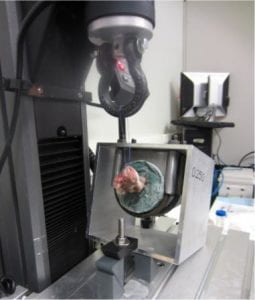
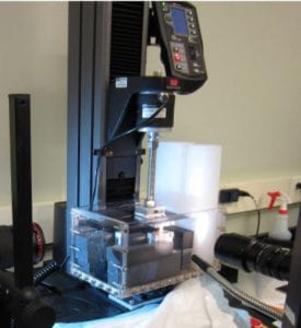
Versatile and easy to use test frame for uniaxial tension or compression with small–medium loads. Ideal for static and slowly varying tests. More often than not this is the tester you should use.
- Displacement range: ~ 1 m
- Displacement rate: arbitrarily slow to ~ 10 mm/s
- Load cells: 10 N, 100 N, 1 kN.
- Bath for testing in fluid
- Single-column test frame provides ample working space.
- A variety of test fixtures (accessories) are available, including a T-slot table, a wide assortment of tensile grips, and compression platens.
- Often used for uniaxial tension, unconfined compression, confined compression, and 3-point bending.
TA 3510 AT
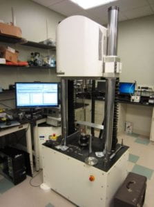
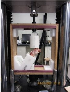
Dynamic uniaxial tension–compression and torsion at moderate loads. Ideal for cyclic testing and high displacement rates. This test frame should be used when the Instron 5943 does not provide adequate performance.
- Displacement range: ± 25 mm. Starting at ~ 0 mm is recommended.
- Displacement rate: 0.025 μm/s to 1.5 m/s, or 0.00001 Hz to 100 Hz.
- Max axial load: 7500 N dynamic, 5300 N static
- Motorized crosshead height adjustment.
- Rotation range: ± 3600° at up to 50 Hz
- Max torque: 49 N·m dynamic, 42 N·m static
- Load cells: 1250 N axial, 7500 N axial, 70 N·m torque
- Test space: 559 mm wide × 2700 mm tall
- Temperature-controlled bath for testing in fluid
- Accessories include MRI-compatible (available at CBBI) locking compression fixtures
TA 3230 AT Series II
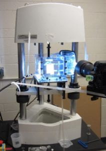 Dynamic uniaxial tension–compression and torsion at small loads. Ideal for cyclic testing and high displacement rates. This test frame should be used when the Instron 5943 does not provide adequate performance.
- Displacement range: ± 12.5 mm. Starting at ~ 0 mm is recommended
- Displacement rate: 0.0065 μm/s to 3.2 m/s, or 0.00001 Hz to 200 Hz
- Max axial load: 450 N dynamic, 320 N static
- Manual crosshead height adjustment with microadjust
- Rotation range: ± 3600° at up to 100 Hz
- Max torque: 5.6 N·m
- Load cells: 10 N axial, 45 N axial, 450 N axial, 0.2 N·m torque, 5.6 N torque
- Test space: 355 mm wide × 417 mm tall (340 mm w/ torsion motor) × 279 mm deep
- Temperature-controlled bath for testing in fluid
- Adapters for Instron test fixtures are available
Microtensile Tester
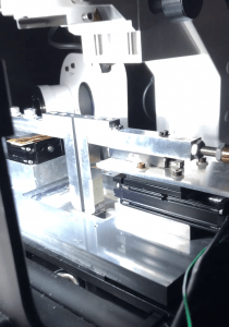
Uniaxial tension tester design for small forces and simultaneous imaging of the specimen with a confocal or multiphoton microscope. Also useful if it is important that the specimen remain horizontal.
- Compatible with Zeiss 880 microscope at APB
- Displacement: up to 50 mm inter-grip distance
- Displacement rate: arbitrarily slow to ~ 20 mm/s
- Load cells: 10 N, 45 N
- Integrated bath with glass coverslip viewing window for inverted microscopy
Biaxial Tension Tester
 Biaxial tension tester for more complete characterization of mechanical properties than is possible through uniaxial testing. It is often impossible to produce an accurate predictive model for an anisotropic material from uniaxial test data alone.
- Displacement range: ~ 10 cm
- Displacement rate: arbitrarily slow to ~ 1 mm/s
- Load cells: 20 N
- Integrated fiducial marker tracking using a camera mounted above the specimen. Fiducial marker strain is calculated in real time and may be used as the test’s controlled variable.
- 4-motor design so the specimen remains centered in the camera’s field of view.
- Integrated rotating polarizers for polarimetry of birefringent materials, for example to measure collagen fiber alignment in tendon or ligament.
- The actuators are connected to the specimen using long sutures.
- Bath for testing in fluid.
NAC HX-7 High-Speed Camera
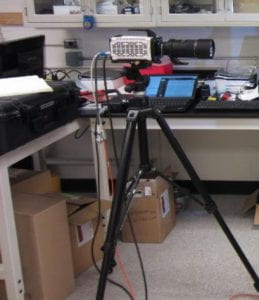 High-resolution and high-framerate video camera. This camera configuration maximizes resolution at the expense of exhibiting a mid-range framerate. Generally this is the correct trade-off for tests of biological tissue.
- 2560 × 1920 px @ 850 fps
- 1920 × 1080 px @ 2000 fps
- Resolution may be sacrificed further for greater framerate.
- Macro (1× magnification) capable: 30 mm × 20 mm field of view at ~ 40 cm working distance.
- 32 GB memory buffer
Scanning Laser Interferometer
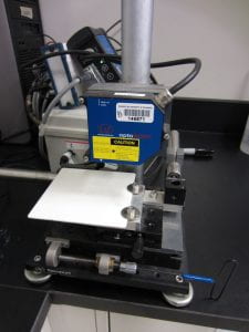 The scanning laser interferometer measures the height profile (i.e., cross-sectional area) of a specimen without contact, which is helpful when the specimen is too soft for accurate micrometer measurements. The specimen is placed flat on an x–y stage and the laser interferometer records a height profile as the x–y stage is traversed.
Zeiss AxioLab A.1 Pol
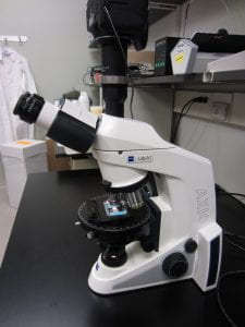 Transmitted light microscope with linear and circular polarizing filters and a rotating x–y stage. Primarily intended for polarimetry and other transmitted light imaging of slide-mounted sections. Includes a camera for image capture.
Zeiss SV-6 Stereomicroscope
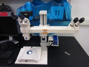 Stereomicroscope for fine dissection and whole-mount imaging of specimens before or after a test. Includes a camera for image capture.
Orthoscan Mini C-Arm Fluoroscope
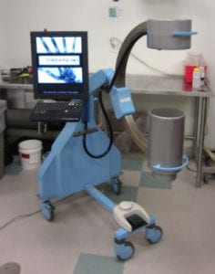 Continuous low-dose x-ray imaging. Still photos may be triggered using a footswitch. Particularly useful for guided injection or minimally invasive surgery and joint alignment.
Leica SM2400 Sledge Microtome with freezing stage
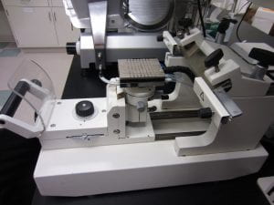 The freezing stage allows soft tissues to be cut to precise dimensions, with parallel faces and thus uniform thickness. The freezing stage is somewhat larger than typically available in a cryotome rotary microtome.
Deli Slicer

A deli slicer is available for rapid rough cutting of soft tissue, which is usually cut while semi-frozen and embedded in a cellulose gel. For tough tissues, the resulting slices are somewhat uneven in thickness, but better than using a vibratome or cutting by hand. This is a quicker but lesser-quality alternative to the Leica SM2400 freezing stage microtome.
Vic-2D Digital Image Correlation Software
 Measure 2D strain fields from a time lapse image series of a specimen with a speckle-patterned surface. Typically used for analysis of uniaxial or biaxial tension test data.
TA Instruments ElectroForce 400N TestBench
 Dynamic uniaxial tension–compression in a horizontal configuration, up to 400 N. Ideal for cyclic testing and high displacement rates. Convenient for small animal work.
- Displacement range: ± 6.5 mm. Starting at ~ 0 mm is recommended
- Cycle frequency up to 100 Hz
- Max axial load 400 N dynamic, ~ 280 N static.
- Load cells: 400 N, 225 N, 22 N (PID only), 5 N (PID only)
- Available test space up to 21.9” (without load cell)
- Adapters allow use of Instron 12 mm socket fixtures
Aurora Scientific 1300A 3-in-1 Whole Animal Muscle Stimulator for Mice
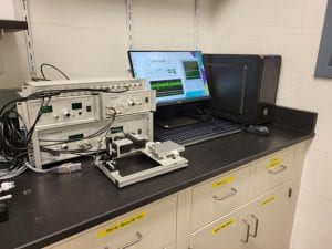
The 3-in1 whole animal muscle stimulator is a high performing, precise test system to test tensile and other mechanical properties of muscle. This system combines in situ, in vivo and in vitro muscle testing using one platform to study muscle physiology in rodent (mouse) subjects. Features include a precision force transducer for in situ tests, force transducer with plate for in vivo tests, and 25mL horizontal bath for in vitro tests. Peak forces range from 0.5 to 10N.
Preclinical Model Lab
Christina Stinger, BS, CVT
Christina Stinger, is a certified Veterinary Technician in the Office of Lab Animal Medicine (OLAM). For the first part of her career, Ms. Stinger worked in exotics and small animal practice and transitioned to lab animal medicine in 2008, as a vet tech and vivarium manager at the Drexel University College of Medicine, in Philadelphia.
In the spring of 2021 she helped design and launch BMEG667: Research Techniques for Preclinical Analysis in Rodents, a lab based course which teaches animal use techniques, research protocol design and implementation. She currently provides services in anesthesia, surgery and imaging for the CPAs core. She is looking forward to growing with the department and helping researchers navigate their studies within the guidelines of animal use and care.
Services offered:
- Analysis assistance
- Assay/Protocol Development
- Assist in Preclinical Experiments
- Conduct Pilot Preclinical Experiments
- Consultation/Training Fee
- Equipment education
- Execution of Preclinical Experiments
- Project development
- Sample processing
- Techniques education
Veterinary/Pre-clinical Analysis Equipment:
Port-X IV Portable Dental X-Ray System
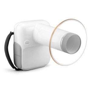 The Portable X-ray system is a devices for photographing structures for accurate diagnosis. This instrument provides high quality images that can be integrated with a mobile app while also reducing radiation exposure. This x-ray system is portable and can easily by operated with one hand.
- Easy to Use
- Wireless
- Mobile App
- Light weight
- Patient Management
- Image Viewer
- Direct Connection with Sensor
Panlab Infrared (IR) Actimeter
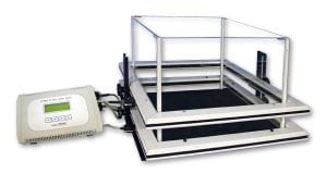
The Panlab (Infrared) Actimeter is an IR based system that allows the study of spontaneous locomotion activity in rodents. The system is composed of a 2 dimensional square frame, frame support and controller. Each frame contains 16×16 infrared beams for optimal subject detection. Each frame can be used to evaluate general activity, locomotion movements or rearings and explorations. The IR actimeter is compatible with mice and rats.
