Multiscale Assessments Research Core (DCMR Research Core)
Multimodal Intravital Imaging Lab
Mary Boggs, PhD
Dr. Mary Boggs completed her PhD at the University of Delaware in the Department of Biological Sciences under the direction of Dr. Randall Duncan from Biological Sciences and Dr. Tom Beebe, Jr. from the Department of Chemistry and Biochemistry in 2011. Her graduate work focused on developing bone/nerve cell co-culturing methods and identifying mechanisms of cell-cell communication between these cells. Post graduation, she stayed on as a Research Associate in the Duncan Lab and was involved in a variety of projects aimed to understand biological molecules involved in bone and cartilage responses to mechanical forces at both the cell and organismal level. Before joining the DCMR, Dr. Boggs served as the Associate Director of DBI Research at the Delaware Biotechnology Institute. In this role she provided K-12 STEM education opportunities and training and provided oversight to the shared instrument facilities at the DBI. Additionally, she served as an Associate Director for the Chemistry/Biology Interface Program where she provided insight and advice to student progress and engaged students in outreach activities and resilience training. As the Lead Scientist for the DCMR, Dr. Boggs is excited to provide her expertise and technical support for those using the DCMR Core Instrumentation.
")
Facilities and Resources:
The Multimodal Preclinical Imaging Laboratory includes several spaces on central UD campus including equipment spaces in rm 244 in the Center for Biomedical and Brain Imaging (CBBI) and 203 Spencer Lab, and offline data analysis space in 115 Wolf Hall. The laboratory provides various resources for high-resolution in vivo, ex vivo and/or in vitro imaging including IVIS, microCT, photoacoustic ultrasound, confocal microscopy, and ocular imaging. Offline analysis includes 3 high-end workstations with software for data analysis associated with laboratory equipment. The DCMR Lead Scientist is available to provide expertise and training on equipment within the facilities.
Services offered:
- Analysis Assistance
- Assay/Protocol Development
- Consultation/Training
- Project Development
Equipment:
Agilent BioTek Cytation C10 Confocal Imaging Reader
 The Cytation C10 is a confocal imaging reader that combines automated confocal and widefield microscopy with multimode plate reading. The system is outfitted with a Hamamatsu Orca sCMOS, 16-bit grayscale camera for use in confocal and widefield imaging. For confocal imaging, users can select between 60μm and 40μm Nipkow spinning discs as well as 6 different laser lines. Available features for confocal and widefield imaging include CY5, DAPI, GFP, RFP, YFP (widefield only) and TRITC filters as well as 1.25, 4, 10, 20, 40 and 60x objectives (confocal is only available for 20, 40 and 60x). The system provides imaging modes including single and multi-color, time lapse, montage, z-stacking and z-stack montage. Detection modes include UV-Vis absorbance, fluorescence intensity and luminescence. The Cytation C10 accepts multi-well microplates ranging from 6 to 1536 wells for imaging and 6 to 384 wells for detection. Other supported labware includes microscope slides, petri and cell culture dishes, cell culture T25 flasks and hemocytometer. To maintain proper environment during time lapse or kinetic studies, temperature can be controlled from room temperature up to 45ºC and CO2 can be controlled from 0-20%.
Location: CBBI (Center for Biomedical and Brain Imaging) 244A
Contact person: Dr. Mary Boggs
Brüker SkyScan 1276 Micro-CT
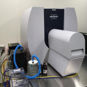 The SkyScan 1276 is a high performance, stand-alone, fast, desktop in vivo micro-CT with continuously variable magnification for scanning small laboratory animals (mice, rats, hamster, etc.) and biological samples.
Description: The Brüker SkyScan 1276 Micro-CT features a combination of high resolution, big image size, round/spiral scanning, reconstruction, and low dose imaging. The image field of view (up to 80 mm wide and more than 300 mm long) allows full body mouse and rat scanning. The variable magnification allows scanning bone and tissue samples with high spatial resolution down to a 2.8 µm pixel size. Variable X-ray energy combined with a range of filters ensures optimal image quality for diverse research applications from lung tissue to bone with metal implants. Further, the SkyScan 1276 in vivo micro-CT administers low radiation dose to the animals allowing multiple scans in longitudinal pre-clinical studies without the risk of unwanted radiation-induced side effects. The system can perform scanning with continuous gantry rotation and in step- and-shoot mode with the fastest scanning cycle of 3.9 sec.
The SkyScan 1276 allows reconstructing non-invasively any cross section(s) through the animal body with possibilities to convert reconstructed datasets into realistic 3D-images and to calculate internal morphological parameters including specific bone structural parameters.
Features:
- 3.9 second shortest scanning cycle
- 2.8 micron highest nominal resolution
- Maintenance-free 20-100 kV micro-focus X-ray source, automatic 6-position filter changer
- 11 Mp cooled X-ray camera
- Continuously variable magnification with smallest pixel size of 2.8 micron
- Possibility to resolve object details of 5-6µm with more than 10% contrast
- Scanning in circular or spiral (helical) trajectory
- GPU-accelerated and world’s fastest InstaRecon® 3D reconstruction
- Full body mouse and rat scanning, 80mm scanning diameter, >300mm scanning length,
- Step-and-shoot and continuous gantry rotation with shortest scanning cycle of 3.9 sec.
- Integrated physiological monitoring (breathing, movement detection, ECG) for time-resolved 4D microtomography
- Software for 2D/3D image analysis, bone morphology and realistic visualization
- On-screen dose meter to estimate accumulated dose and dose rate
- Export results to 3D printers to create magnified physical copy of scanned objects
Cellink BioX6 Bioprinter
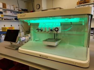 The BioX6 Bioprinter is a high-throughput bioprinter for cell culturing and biofabrication. The BioX6 provides 6 printheads to print more complex structure and physiologically relevant models. The printhead temperatures can range from 4-250ºC with a printbed temperature of 4 to 65ºC allowing for precise control when working with temperature-sensitive materials. The bioprinter is enclosed in a benchtop biosafety cabinet with a HEPA filter and 4 build-in UV modules to maintain sterility around the print area.
Location: CBBI (Center for Biomedical and Brain Imaging) 244 Main
Contact person: Dr. Mary Boggs
FujiFilm VisualSonics Vevo F2 LAZR-X Photoacoustic Ultrasound
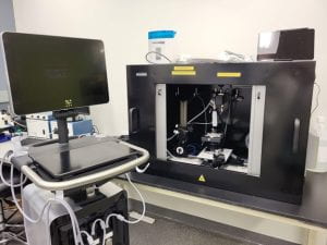 The Vevo F2 LAZR-X Photoacoustic Ultrasound is a multimodal imaging system that provides high resolution, high frequency ultrasound imaging of rodent models including mouse and rat. Ultrasound imaging depths can be up to 9cm with lower frequency transducers, while resolution can be increased (down to 30μm) using higher-frequency transducers. The added LAZR-X allows users to visualize real-time oxygen saturation and molecular imaging, co-registered with high resolution anatomy. Available ultrasound transducers include UHF29x, UHF46x and UHF71x. Photoacoustic imaging is available using the UHF29x and the UHF46x transducers.
Location: CBBI (Center for Biomedical and Brain Imaging) 244A
Contact Person: Dr. Mary Boggs
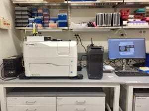
IVIS Lumina III In Vivo Imaging System
 The IVIS ® Lumina Series III brings together years of leading optical imaging technologies into one easy to use and exquisitely sensitive bench-top system. The IVIS Lumina III is capable of imaging both fluorescent and bioluminescent reporters. The system is equipped with up to 26 filter sets that can be used to image reporters that emit from green to near-infrared. Superior spectral unmixing can be achieved by Lumina III’s optional high resolution short cut off filters.
Features:
- Exquisite sensitivity in bioluminescence
- Full fluorescence tunability through the NIR spectrum
- Compute Pure Spectrum spectral unmixing for ultimate fluorescence sensitivity
- Dedicated XGI-8 Gas Anesthesia System
Location: CBBI (Center for Biomedical and Brain Imaging) 244A
Contact person: Dr. Mary Boggs
Phoenix MICRON IV
 Description: Coming soon
Location: Life Science Research Facility (LSRF) 106A
Contact person: Dr. Mary Boggs
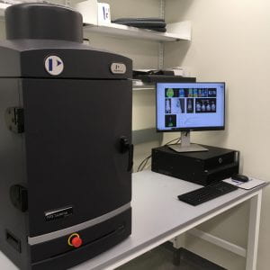
Scanco Medicine 35 micro CT
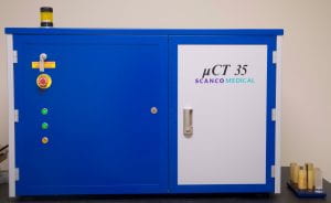
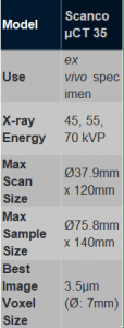 The Scanco 35 microCT system is used to exam diverse materials including bone and other hard tissues as well as medical devices. This system can be used to assess trabecular bone structure from tibia, femor and vertebrae harvested from rats and mice.
Location: Spencer Lab 203
Contact person: Dr. Mary Boggs
Scintica Osteosys iNSiGHT DXA
 Description: coming soon
Location: Life Science Research Facility (LSRF) 208
Contact person: Dr. Mary Boggs and Faysal Haque
Wacom Cintiq Pro 24 touch

A touch tablet with wireless pen for precision data segmentation.
Location: Wolf Hall 115A
Contact person: Dr. Mary Boggs
Workstations
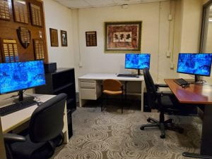 The DCMR supports a set of computational workstations in 203 Spencer Lab (2 units), 244 CBBI (1 unit) and 115 Wolf hall (3 units). Each set of workstations provide software for offline data analysis for equipment related to multimodal imaging including the Bruker Skyscan softwares and Vevo Lab, and other softwares such as ImageJ, MATLAB, Python and the Materialise Mimics software suite for segmentation and FEA.
Location: Wolf Hall 115A
Contact person: Dr. Mary Boggs