Equipment:
Multimodal Animal Imaging Center
Brüker SkyScan 1276 Micro-CT
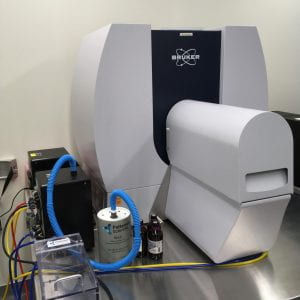 The SkyScan 1276 is a high performance, stand-alone, fast, desktop in vivo micro-CT with continuously variable magnification for scanning small laboratory animals (mice, rats, hamster, etc.) and biological samples.
Description: The Brüker SkyScan 1276 Micro-CT features a combination of high resolution, big image size, round/spiral scanning, reconstruction, and low dose imaging. The image field of view (up to 80 mm wide and more than 300 mm long) allows full body mouse and rat scanning. The variable magnification allows scanning bone and tissue samples with high spatial resolution down to a 2.8 µm pixel size. Variable X-ray energy combined with a range of filters ensures optimal image quality for diverse research applications from lung tissue to bone with metal implants. Further, the SkyScan 1276 in vivo micro-CT administers low radiation dose to the animals allowing multiple scans in longitudinal pre-clinical studies without the risk of unwanted radiation-induced side effects. The system can perform scanning with continuous gantry rotation and in step- and-shoot mode with the fastest scanning cycle of 3.9 sec.
The SkyScan 1276 allows reconstructing non-invasively any cross section(s) through the animal body with possibilities to convert reconstructed datasets into realistic 3D-images and to calculate internal morphological parameters including specific bone structural parameters.
Features:
- 3.9 second shortest scanning cycle
- 2.8 micron highest nominal resolution
- Maintenance-free 20-100 kV micro-focus X-ray source, automatic 6-position filter changer
- 11 Mp cooled X-ray camera
- Continuously variable magnification with smallest pixel size of 2.8 micron
- Possibility to resolve object details of 5-6µm with more than 10% contrast
- Scanning in circular or spiral (helical) trajectory
- GPU-accelerated and world’s fastest InstaRecon® 3D reconstruction
- Full body mouse and rat scanning, 80mm scanning diameter, >300mm scanning length,
- Step-and-shoot and continuous gantry rotation with shortest scanning cycle of 3.9 sec.
- Integrated physiological monitoring (breathing, movement detection, ECG) for time-resolved 4D microtomography
- Software for 2D/3D image analysis, bone morphology and realistic visualization
- On-screen dose meter to estimate accumulated dose and dose rate
- Export results to 3D printers to create magnified physical copy of scanned objects
IVIS Lumina III In Vivo Imaging System
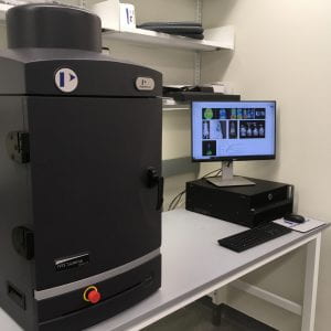 The IVIS ® Lumina Series III brings together years of leading optical imaging technologies into one easy to use and exquisitely sensitive bench-top system. The IVIS Lumina III is capable of imaging both fluorescent and bioluminescent reporters. The system is equipped with up to 26 filter sets that can be used to image reporters that emit from green to near-infrared. Superior spectral unmixing can be achieved by Lumina III’s optional high resolution short cut off filters.
Features:
- Exquisite sensitivity in bioluminescence
- Full fluorescence tunability through the NIR spectrum
- Compute Pure Spectrum spectral unmixing for ultimate fluorescence sensitivity
- Dedicated XGI-8 Gas Anesthesia System
Location: CBBI (Center for Biomedical and Brain Imaging) 244A
Contact person: Dr. Mary Boggs
Scanco Medicine 35 micro CT
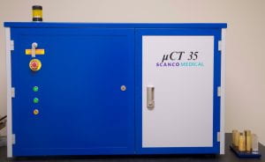
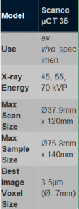 The Scanco 35 microCT system is used to exam diverse materials including bone and other hard tissues as well as medical devices. This system can be used to assess trabecular bone structure from tibia, femor and vertebrae harvested from rats and mice.
Histology Lab
Leica RM2155 Rotary Microtome
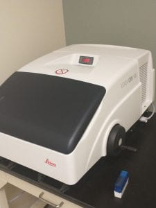
 The Leica RM2155 is a reliable rotary microtome designed for fully motorized sectioning of both paraffin and resin-embedded specimens. Offering a broad application spectrum in histology laboratories, the RM2155 also has applications in industrial materials and quality control. The Leica microtome’s two-in-one design concept allows motorized as well as manual sectioning, and provides reproducible quality sections.
The RM2155 sectioning speed can be adjusted to cut hard materials as well as paraffin-embedded specimens at high throughput without affecting sectioning quality. This microtome is designed for users who require a fully motorized/electronic microtome for their laboratory needs, making it an ideal instrument for fundamental sectioning of standard and non-standard materials.
The fully motorized Leica RM2155 microtome can cut sections from 0.5 to 60 microns with an automatic motorized coarse feed as well as an adjustable hand wheel for speed control and selectable cutting modes. The knife holder comes with new clamping plates, ensuring the user accurate, non-erratic sectioning.
Leica CM 1100 Tabletop Cryostat
The Leica CM1100 is a portable compact cryostat for rapid freezing and manual sectioning of tissue specimens.
Features
- Portable bench-top cryostat
- Compact unit, providing maximum ease of use at a total weight of 50 kg
- Spacious, easy-to-clean cryochamber made out of stainless steel
- CE disposable blade holder with lateral adjustment and glass anti-roll plate
- Quick-freeze shelf
- Two section waste trays
- Microtome with specimen cylinder slot cover – ideal for spray disinfection
- Automatic and manual hot gas defrosting
- CFC-free refrigerants and insulating foams.
- Connection for 12-volt car battery
Technical Specifications:
- Section thickness adjustment: 0 to 20 μm
- Maximum specimen size: 25 mm ø
- Total horizontal specimen advance: 10 mm
- Vertical specimen stroke: 46 mm
- Dimensions (W x H x D): 570 x 380 x 777 mm
- Dimensions cryocabinet (W x H x D): 265 x 220 x 400 mm
- Weight (including microtome): 110 lbs.
Zeiss Upright Light Microscope
Description coming soon
Printer ESPO Thermal Transfer Slide Blue- coming soon
Description coming soon
Pi Single Hopper Plus Cassette Printer- coming soon
Description coming soon
HistoDream Embedding Workstation- coming soon
Description coming soon
Tissue-Tek Film 4740 Coverslipper- coming soon
Description coming soon
Leica ASP6025 S Tissue Processor- coming soon
Description coming soon
Leica Autostainer XL (ST5010) - coming soon
Description coming soon
Avantik QS12 Cryostat- coming soon
Description coming soon
Leica RM2125 Microtome- coming soon
Description coming soon
IsoMet Low Speed Saw- coming soon
Description coming soon
Tissue-Tek Genie Advanced Staining System- coming soon
Description coming soon
Veterinary/Pre-clinical Analysis
Port-X IV Portable Dental X-Ray System
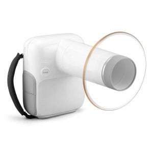 The Portable X-ray system is a devices for photographing structures for accurate diagnosis. This instrument provides high quality images that can be integrated with a mobile app while also reducing radiation exposure. This x-ray system is portable and can easily by operated with one hand.
- Easy to Use
- Wireless
- Mobile App
- Light weight
- Patient Management
- Image Viewer
- Direct Connection with Sensor
Mechanical Loading and Measurement Lab
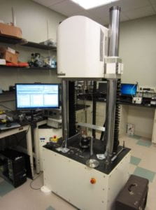
Instron 5943
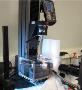
Versatile and easy to use test frame for uniaxial tension or compression with small–medium loads. Ideal for static and slowly varying tests. More often than not this is the tester you should use.
- Displacement range: ~ 1 m
- Displacement rate: arbitrarily slow to ~ 10 mm/s
- Load cells: 10 N, 100 N, 1 kN.
- Bath for testing in fluid
- Single-column test frame provides ample working space.
- A variety of test fixtures (accessories) are available, including a T-slot table, a wide assortment of tensile grips, and compression platens.
- Often used for uniaxial tension, unconfined compression, confined compression, and 3-point bending.
TA 3510 AT
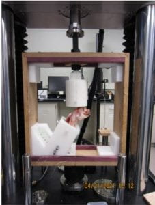
Dynamic uniaxial tension–compression and torsion at moderate loads. Ideal for cyclic testing and high displacement rates. This test frame should be used when the Instron 5943 does not provide adequate performance.
- Displacement range: ± 25 mm. Starting at ~ 0 mm is recommended.
- Displacement rate: 0.025 μm/s to 1.5 m/s, or 0.00001 Hz to 100 Hz.
- Max axial load: 7500 N dynamic, 5300 N static
- Motorized crosshead height adjustment.
- Rotation range: ± 3600° at up to 50 Hz
- Max torque: 49 N·m dynamic, 42 N·m static
- Load cells: 1250 N axial, 7500 N axial, 70 N·m torque
- Test space: 559 mm wide × 2700 mm tall
- Temperature-controlled bath for testing in fluid
- Accessories include MRI-compatible (available at CBBI) locking compression fixtures
TA 3230 AT Series II
 Dynamic uniaxial tension–compression and torsion at small loads. Ideal for cyclic testing and high displacement rates. This test frame should be used when the Instron 5943 does not provide adequate performance.
- Displacement range: ± 12.5 mm. Starting at ~ 0 mm is recommended
- Displacement rate: 0.0065 μm/s to 3.2 m/s, or 0.00001 Hz to 200 Hz
- Max axial load: 450 N dynamic, 320 N static
- Manual crosshead height adjustment with microadjust
- Rotation range: ± 3600° at up to 100 Hz
- Max torque: 5.6 N·m
- Load cells: 10 N axial, 45 N axial, 450 N axial, 0.2 N·m torque, 5.6 N torque
- Test space: 355 mm wide × 417 mm tall (340 mm w/ torsion motor) × 279 mm deep
- Temperature-controlled bath for testing in fluid
- Adapters for Instron test fixtures are available
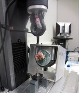
Microtensile Tester
Uniaxial tension tester design for small forces and simultaneous imaging of the specimen with a confocal or multiphoton microscope. Also useful if it is important that the specimen remain horizontal.
- Compatible with Zeiss 880 microscope at APB
- Displacement: up to 50 mm inter-grip distance
- Displacement rate: arbitrarily slow to ~ 20 mm/s
- Load cells: 10 N, 45 N
- Integrated bath with glass coverslip viewing window for inverted microscopy
Biaxial Tension Tester
 Biaxial tension tester for more complete characterization of mechanical properties than is possible through uniaxial testing. It is often impossible to produce an accurate predictive model for an anisotropic material from uniaxial test data alone.
- Displacement range: ~ 10 cm
- Displacement rate: arbitrarily slow to ~ 1 mm/s
- Load cells: 20 N
- Integrated fiducial marker tracking using a camera mounted above the specimen. Fiducial marker strain is calculated in real time and may be used as the test’s controlled variable.
- 4-motor design so the specimen remains centered in the camera’s field of view.
- Integrated rotating polarizers for polarimetry of birefringent materials, for example to measure collagen fiber alignment in tendon or ligament.
- The actuators are connected to the specimen using long sutures.
- Bath for testing in fluid.
NAC HX-7 High-Speed Camera
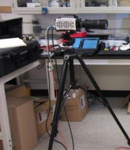 High-resolution and high-framerate video camera. This camera configuration maximizes resolution at the expense of exhibiting a mid-range framerate. Generally this is the correct trade-off for tests of biological tissue.
- 2560 × 1920 px @ 850 fps
- 1920 × 1080 px @ 2000 fps
- Resolution may be sacrificed further for greater framerate.
- Macro (1× magnification) capable: 30 mm × 20 mm field of view at ~ 40 cm working distance.
- 32 GB memory buffer
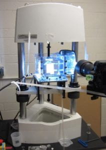
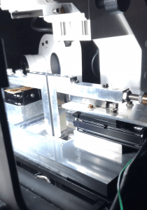
Scanning Laser Interferometer
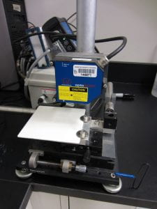 The scanning laser interferometer measures the height profile (i.e., cross-sectional area) of a specimen without contact, which is helpful when the specimen is too soft for accurate micrometer measurements. The specimen is placed flat on an x–y stage and the laser interferometer records a height profile as the x–y stage is traversed.
Zeiss AxioLab A.1 Pol
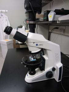 Transmitted light microscope with linear and circular polarizing filters and a rotating x–y stage. Primarily intended for polarimetry and other transmitted light imaging of slide-mounted sections. Includes a camera for image capture.
Zeiss SV-6 Stereomicroscope
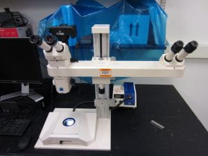 Stereomicroscope for fine dissection and whole-mount imaging of specimens before or after a test. Includes a camera for image capture.
Orthoscan Mini C-Arm Fluoroscope
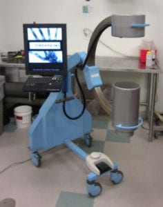 Continuous low-dose x-ray imaging. Still photos may be triggered using a footswitch. Particularly useful for guided injection or minimally invasive surgery and joint alignment.
Leica SM2400 Sledge Microtome with freezing stage
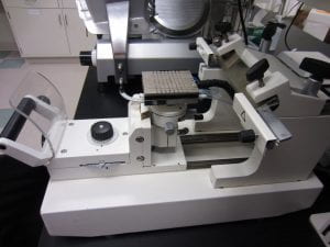 The freezing stage allows soft tissues to be cut to precise dimensions, with parallel faces and thus uniform thickness. The freezing stage is somewhat larger than typically available in a cryotome rotary microtome.
Deli Slicer

A deli slicer is available for rapid rough cutting of soft tissue, which is usually cut while semi-frozen and embedded in a cellulose gel. For tough tissues, the resulting slices are somewhat uneven in thickness, but better than using a vibratome or cutting by hand. This is a quicker but lesser-quality alternative to the Leica SM2400 freezing stage microtome.
Vic-2D Digital Image Correlation Software
 Measure 2D strain fields from a time lapse image series of a specimen with a speckle-patterned surface. Typically used for analysis of uniaxial or biaxial tension test data.

About
The University of Delaware Bio-Imaging Center is a multi-user microscopy facility that houses state-of-the-art equipment. Our staff has technical expertise across the gamut of microscopy platforms and methodologies. With a special emphasis on life sciences and soft materials, we are able to address the broad imaging needs of the university, non-profit and industrial research community. The center is open to all academic researchers and industrial clients on a fee-for-service basis.
Location
The center is housed at the Ammon Pinizzotto Biopharmaceutical Innovation Center (Suite 141), Center for Biomedical and Brain Imaging (CBBI 244), and the Department of Biological Sciences, 267 and 304 Wolf Hall.